He woke up in an unfamiliar room to a nurse asking, “Who is the president of the United States?”
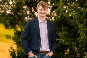
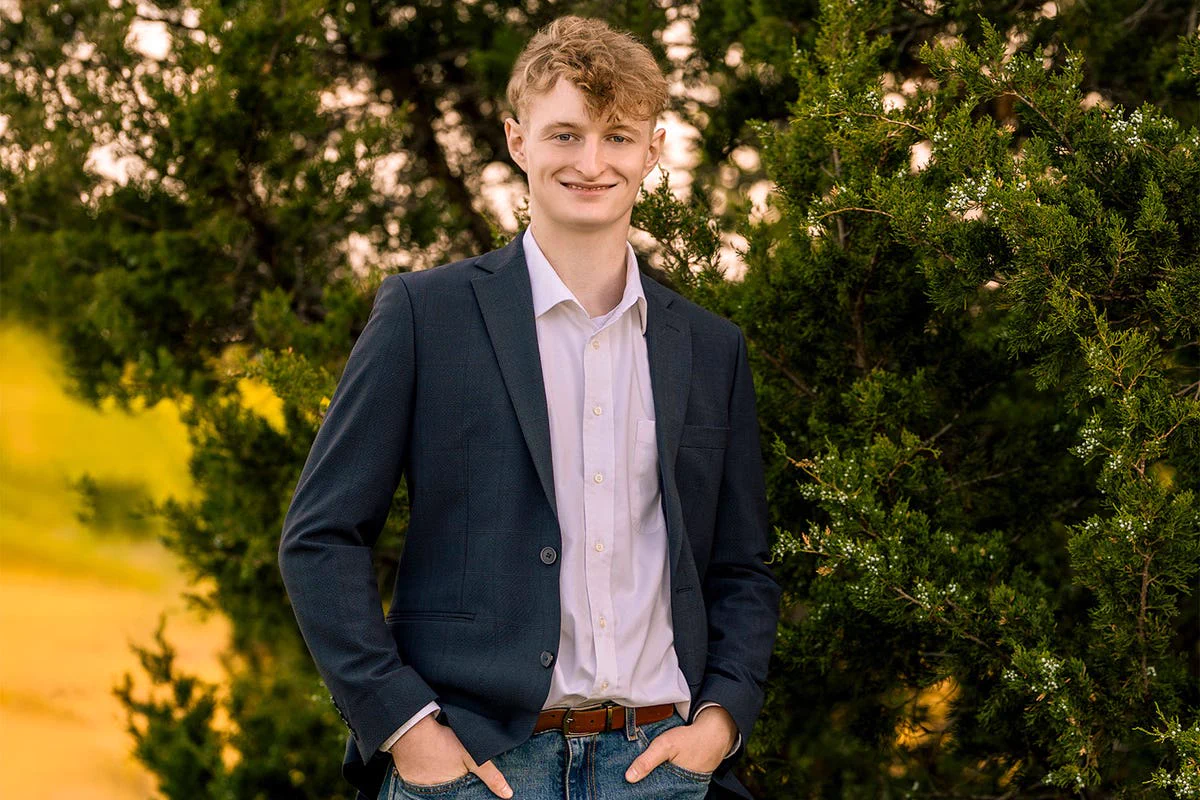
The last thing 17-year-old Dylan Lindley of Plano, Texas, remembered was hanging out at a skate park with buddies and then going to sleep at a friend’s house early the next morning. Now, he was lying in a hospital bed, surrounded by his parents and older brother, and utterly bewildered about what was going on.
As Dylan later learned, he’d had a seizure shortly after falling asleep. Fortunately, his friend saw what was happening and told his father, who immediately called Dylan’s house. Minutes later, Dylan’s dad and brother arrived and took him to a local emergency room.
An initial CT scan showed a blurry mass on the left side of his brain, which an MRI scan later confirmed was a brain arteriovenous malformation (AVM). This rare condition, marked by a tangle of abnormal blood vessels, can be fatal.
The diagnosis started Dylan on a journey that culminated in a cure at the Mass General Brigham Neuroscience Institute.
Finding one of the world’s foremost AVM experts
Dylan had a grade 4 AVM. The mass was located near eloquent structures of the brain (critical areas that control language, speech, reading, and motor function). Disrupting any of these regions could cause severe, permanent neurological damage. For this reason, many centers deem AVMs like Dylan’s too high risk for surgery.
A local neurologist said it would be impossible to remove Dylan’s AVM surgically. Instead, he proposed treating it with radiation therapy.
“He said there was a 50/50 chance the radiation would work and we wouldn’t know for three years whether it had been successful,” Dylan said. “In the meantime, the AVM could rupture and kill me.”
Dylan and his family didn’t like those odds. His mother, Haley, started researching other options. Several centers either didn’t offer surgery or couldn’t review Dylan’s case in a timely manner. Then she discovered Nirav Patel, MD, a neurosurgeon with the Neuroscience Institute.
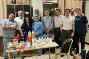
Dr. Patel has devoted his career to the treatment of complex brain AVMs. Over the past 15 years, he has built a multidisciplinary team of neurosurgeons, neurologists, neuroradiologists, radiation oncologists, neurointensivists, anesthesiologists, rehabilitation specialists, and nurses dedicated to caring for patients with these rare lesions. Their specialized expertise and experience draw patients with complex AVMs from across the United States and beyond.
About a month after his seizure, Dylan and his parents had a video call with Dr. Patel.
Checking all the boxes
At the Neuroscience Institute, Dylan underwent two types of studies:
- Functional MRI (fMRI), which looks at blood flow to detect activity in specific areas of the brain
- Angiogram, which creates detailed images of blood vessels in the brain
When considering how to treat an AVM, Dr. Patel asks three key questions. First, are he and his team capable of doing the case? Based on the results of the imaging studies, he believed he could extract Dylan’s AVM safely.
Second, should he recommend surgery? This means determining whether a) surgery makes sense for the individual patient, b) it aligns with the patient’s goals, and c) the potential benefits outweigh the risks. The patient’s age is an important factor here. For example, it might not be wise to put an 82-year-old through the stress of an operation. But for an otherwise healthy 17-year-old with his whole life in front of him, Dr. Patel thought surgery was a viable option.
Finally, is the patient totally on board with proceeding? Dr. Patel laid out the many risks to gauge Dylan’s commitment.
“I told him, ‘Worst-case scenario, you could end up paralyzed or unable to talk, understand words, or read. You may not be able to finish high school, and you may need rehabilitation for six months,’” Dr. Patel said. “He looked me in the eye and said, ‘Let me tell you, I’m a wrestler. Every time I get pinned down, I fight to get back up. I’m going to be able to do this no matter what.’
“No patient can truly know what it’s like to experience a serious complication before it happens. But a patient’s commitment to pursuing a cure—and their determination to work through whatever recovery may require—does matter.”
The benefits of an arterial-first strategy
A few weeks later, Dylan was lying in a bed at Brigham and Women’s Hospital waiting to be taken into the operating room.
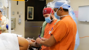
Before surgery, Dr. Patel combined Dylan’s MRI, fMRI, and angiogram into a detailed three-dimensional roadmap of the AVM and the surrounding areas of the brain responsible for movement, language, and other critical functions. During the operation, that roadmap was integrated into the surgical microscope, helping guide the team safely through the procedure.
Using the microscope and tiny instruments, Dr. Patel meticulously went about his work. Typically, neurosurgeons gradually work around an AVM, encountering arteries feeding blood to the malformation along the way.
In contrast, Dr. Patel first identified and disconnected nearly all of the feeding arteries before removing the AVM. This “arterial-first” strategy:
- Helps prevent the AVM from bleeding into the eloquent structures, which could cause irreparable damage to the brain
- Softens the nidus (the “nest” at the center of the tangled blood vessels), which makes it easier to remove later
Once the feeding arteries were addressed, Dr. Patel set out to carefully separate the AVM from the surrounding tissue. Throughout the procedure, senior surgical neurophysiologist Mitali Bose, MS, CNIM, and her neuromonitoring team carefully tracked Dr. Patel’s every move, warning him if he was coming too close to any eloquent areas. The team also kept a close eye on Dylan’s motor, visual, and other functions to confirm they weren’t being compromised.
After Dr. Patel believed he had removed the entire AVM, Dylan was taken to another room for an angiogram. The images showed the AVM was completely gone. The surgery had accomplished what everyone had hoped for—a complete cure while preserving the brain functions that mattered most to Dylan’s future.
High-quality postoperative care is crucial
Traditionally, a patient who has had a high-grade AVM extracted is put in a medically induced coma for seven days. During this period, their blood pressure is kept at a low level to reduce the risk of bleeding in the brain.
However, keeping blood pressure low in a young person like Dylan requires sedation (or “twilight sleep”). That carries its own risks, such as pneumonia.
Dr. Patel and neuroanesthesiologist Grace Kim, MD, have developed a special protocol for cases like this. Dr. Kim discovered she could remove the breathing tube from the patient right after the postoperative angiogram—and still safely manage their blood pressure over the ensuing days. As a result, the patient needs to be kept in a coma for a much shorter period.
Dylan was awoken two days after the surgery. He had been warned that he might not be able to move the right side of his body or the right side of his face might be drooping. When he realized he had only mild numbness on his right side, he was pleasantly surprised. His one major problem was aphasia (inability to communicate), which would require speech therapy.
For seven days, Dylan rested and recovered in the Neuroscience Intensive Care Unit (neuro ICU). As Dr. Patel pointed out, the unit’s nurses play a critical role in managing the patient’s blood pressure and ensuring the enlarged arteries in the brain shrink to a normal size.
“We’re fortunate to have neuro ICU nurses who know the postoperative protocol and have the real-world experience to handle these very complex cases,” Dr. Patel said.
Extensive therapy at Spaulding Rehabilitation
After one week in the neuro ICU, Dylan was transferred to Spaulding Rehabilitation. Every day, he had separate sessions with physical, occupational, and speech therapists. “Spaulding was where I really started getting physically and mentally better,” he said.
Dylan ended up spending two weeks at Spaulding and another week at a local hotel with his family. By the time they flew home, he had been cleared to walk without a walker.
“The right side of my body was still kind of numb, and I still messed up words frequently. Other than that, I felt great physically,” he said.
Back in Plano, Dylan went for physical therapy for a short period. Speech therapy was a more pressing need, as he was still struggling with speaking. With a lot of hard work, those skills slowly came back.
Three months post-surgery, Dylan began his postponed senior year of high school. About a month later, he rejoined the wrestling team. More milestones followed, culminating in graduation. He was just what he wanted to be again: a typical kid.
Ready to start a new chapter
With the operation almost a year behind him, Dylan, 18, is looking forward to attending Texas’ Blinn College in the fall.
“I can do everything I used to do,” he said. “Everything’s pretty much back to normal.”
Dr. Patel isn’t surprised. “Too often, young people like Dylan are told, ‘You can never get married, you can’t have children, you should avoid contact sports, you should consider a career that doesn’t require much physical activity,’” Dr. Patel said. ”In fact, Dylan can go on and live a normal life. He could play football, fly a plane, scuba dive—he has absolutely no limits from a medical perspective.”
Dylan will return to Boston most every year for follow-up imaging and visits with Dr. Patel and his team. Instead of planning his life around an untreated AVM, he is planning for college, his career, and everything else that comes with being an 18-year-old.
“Dr. Patel saved my life,” Dylan said. “There was no one else who was going to take that AVM out. I owe everything to him and the rest of his team. I think they’re extraordinary.”